APPARATUS (Reagents and equipments):
4-5 clean glass slides, Leishman’s stain, blood lancet, dropper, glass rod, compound microscope, cedar wood oil, buffered water and staining tray.
PRINCIPLE:
A blood film appropriately prepared and stained is seen under the microscope in oil immersion. Different WBCs are seen which are counted in percentage. The total 100 leukocytes are studied, identified and recorded from a blood smear.
PROCEDURE:
- Prepare, stain and examine a blood smear under oil immersion lens.
A. Blood film preparation by Wedge technique.
- Place 3 x 1 inch glass slide on a flat surface (Stationary slide).
- A drop of blood is placed on the writing hand side of the slide.
- It is spreaded by a haemocytometer cover glass by pushing slide forward with smooth and rapid stroke so that the blood will be pulled behind the spreader.
Characteristics of a properly prepared wedge film are as follows:
- The blood should cover at least half length of the slide.
- It should be narrower than the slide width.
- The spread of the blood should be homogenous spread with gradual transition from thick to thin areas i. e. there should be no troughs, holes, streaks, waves and bubbles.
- The end of the film should have feather end or it should have a straight end.
- Bullet shaped preparation of the film have higher accumulation of leucocytes along the sides and tail edge of the film than the straight ended film.
- The film should be thin enough to allow proper fixation. Thick areas appear dark green or gray and are washed off during staining
- The film should have at least 10 low power fields in which half of the RBCs do not ovetlap. The rest of the RBCs may overlap slightly (doublets and triplets).
- The cause of the thick film iseither using a big blood drop or the spread of the film is too quick and at a high angle. The cause of thin film is a blood drop which is too small or the spread of the film is too slow and at a too low angle.
B. Staining of the blood film by Rack Method.
- Place the slide on glass rods overlying a sink or dish that keeps the glass slide in a horizontal position.
- The glass slide is covered with chemically pure methanol (CH3COOH).
- The slide is allowed to dry and the blood film becomes fixed.
- The surface of the blood film is flooded with Leishmann’s stain with the help of a dropper.
- After 10 minutes the film is flushed with the stream of distilled water till all stain is removed.
- The residual stain on the back of the slide is cleaned off with cotton gauze or tissue paper and the specimen is dried in the ‘air.
Criteria for a nicely stained film:
The well stained film should be reddish brown macroscopically. Microscopically, the RBC should be visible salmon pink, the nuclei of the leucocytes should be stained purple blue and the cytoplasm of the platelets should
2. Draw one hundred squares on a paper.
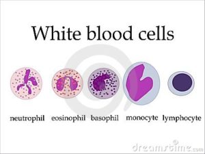
3.The differential cells are counted manually. An area where the film is made quite clear under the low power magnification is selected. The best area for cell counting in a slide is the body of the film and not head or tail. The two drops of cedar wood oil are placed over the slide and the oil immersion lens is shifted in contact with cedar wood oil. Identify various types of leukocyte and enter them by hrst letter (NNeutrophil, EEosinophil, Basophil, Lymphocyte, M-Monocyte) in all 100 squares on the paper. At least 100 WBCs are classrf ed Report the result of 100 cells classified as percentage.
4. Count the cells in a specific sequence to avoid repeated counting. Track method is usually used to count WBCs. (Longitudinal pattern method or lengthwise method is also used) in which cells are countered in consecutive fields in strips or rows from the tail end straight back towards the thitk end of film i.e. cells are counted in one direction and then field is shifted and counting is done in opposite direction. Field is again shifted in the same direction and counting is done in same direction as in the beginning. .
- Percentage of various types of white cell is calculated by counting the different cells in various squares.
Normal Leukocyte count:
1 . Granulocytes.
- Neutrophil
- Eosinophil 1 4%
- Basophil O 1%
-
2.Non-Granulocytes
- Lymphocte 20- 40%
- Monocyte 2- 8% or lilac.
PRECAUTIONS:
- The blood film should be thin and smooth.
- We should not count the same cells twice.
- Same as in preparation of blood smear and identification of various cells.
- The slidesused should be clean.
- Under staining and over staining of the slide should be avoided.
- The lens and slide contact should be guarded carefully to avoid the breakage and also to avoid damage to the slide.
VARIATION
-
a. Neutrophils.
- Neutrophilia: Increase neutrophil count. 1. Severe muscular exercise, pregnancy and food in take.
- Acute pyogenic infections e.g. abscess, boils, tonsillitis, etc.
- Netropenia: Decrease neutrophil count. 1. Depression of bone marrow: Aplastic anemia, bone marrow depressant drug,
e.g. chloramphenicol.
- Typhoid, paratypohoid and malaria.
B Lymphocytes.
a. Lymphocytosis: Increase lymphocyte count.
- In new born and infant lymphocytes are more.
- Chronic infection, e.g. tuberculosis.
- Decreases lymphocytes (lymphopenia) ACTH and steroid therapy.
c.Eosinophil.
Eosinophilia (increase eosinophil count): Allergy, condition like pulmonary eosinophilia, worm infestation, hay fever and urticaria.
- Eosinopenia (decrease eosinophil count): Stress (acute bacterial infection) glucocorticoid administration.
-
Basophils.
- Increase basophil count in chronic myeloid leukemia, small pox and polycythemia.
- Decrease basophil count in acute pyogenic infection.
Monocyte.
Increase monocyte count in infectious mononucleosis, kala-azar and malaria.
DISCLAIMER:
Here i need you attention to some of the important words about this article. This post was share just for educational purpose and to help medical students, post graduates and professors. This Site only educate our visitors about the materials. Medicscenters.com do not share the copyright materials on this site. The Link which is provide as found/ taken from the others websites. we highly encourage our visitors to buy the materials from its original authors. More over we do not store any kind of illegal link in our medicscenter.com server. We always follow the legal DMCA policy. There is no copyright materials share on medicscenter.com, We only offer the materials which are free on internet. This website is educational purpose and if there is any copyright materials against DMCA policy, who want to remove the materials kindly contact us on email [email protected].
Leave a Reply