The purpose of preparation of blood smear (blood film} is to study the morphology of RBCs, differential leukocyte count and reticulocyte count.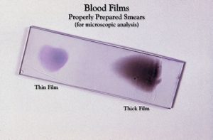
APPARATUS:
4-5 glass slides, compound microscope, pricking needle (blood lancet), spirit swab, cedar wood oil/liquid paraffin, Leishman’s stain, wash bottle, buffered water and staining tray.
Leishman’s Stain:
1.5 gm powder of Leishman’s stain is dissolved in one litre of acetone free methyl alcohol. Leishman’s stain contains two dyes, eosin and methylene blue. Eosin is an acidic dye that stains basic structures like RBC and granules of eosinophil. It is pink or red in colour. Methylene blue is a basic dye that stains acidic structures like nucleus or granules of basophils. It is blue in colour. Acetone free methyl alcohol is a fixative for smear.
- Fixation of smear is because of precipitation of proteins by alcohol which prevent washing off of the film.
- It preserves the cells in what ever chemical and metabolicstate they are at the time of staining.
- Acetone if present will cause shrinkage or even lysis of the cells.
PRINCIPLE:
Blood smear is prepared, stained with Leishman’s stain and cells are identified under oil immersion lens.
PROCEDURE: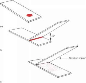
A) Preparation of Blood Smear
- Selection of a spreader: Take one slide a spreader which has smooth edge. It should be done by careful look on the narrow edge of the slide or by moving a thumb smoothly on its edge. But, the slide should be washed with soap and water after touching its edge, to remove grease particles from its edges.
- Take 3-4 clean and dry glass slides and keep them on filter paper or any clean white paper placed on the table.
- Prick the ring finger of left hand with the help of prickling needle under all aseptic conditions (discard the first two drOps of blood from skin puncture site). Specimen of blood can also be taken by venepuncture. Put a small size of blood drop on each glass slide about half centimeter from it’s narrow edge on the right side. Place the blood drop In the same way on other slides also.
- Now put the spreader on left side of blood drop at the angle 45° as shown in Figure 14.
- Give left to right movement to the spreader so that blood comes along the edge of the spreader. Further give side-to-side movement so that blood comes along the whole edge of spreader.
- Now spread the blood by giving smooth, uniform and rapid movement to the spreader upto the left edge of the slide. Repeat the same procedure with rest of the slides.
- Dry the smear by waving the slides in the air for some time.
- Now observe the all smears whether they are satisfactory or not ).
A good smear has following characteristics:
- It is tongue shaped having head, body and tail. Head is the area where blood drop is placed. Body is the area between head and tall. Tail is the last part of the smear with ragged margins.
- It should cover two-thirds of the slide.
- It should not be thick.
- There should not be marks or blank spaces in the smear.
- The dried film should be of brownish yellow colour (Buff coloured).
9. After selecting a good smear with naked eyes focus it under low power of the microscope. The smear should be thin enough to have single layer of cells. There
should not be rouleaux formation or clumping of cells.
B) Staining of Blood Smear
- Leave it for 1-2 minutes for fixation of the smear.
- Add Leisman’s stain drop by drop till it covers whole of the smear. Count the number of drops you have put.
- Place 3-4 good slides horizontally on the stand.
- Add equal number of drops of buffered water (pH 6.8) on the slide. Mix the stain with water by blowing air with the help of a glass tube or with a dropper.
- Wait for 8-10 minutes for staining to complete. During this period in the presence of buffered water staining is taking place because of formation of cations and anions of basic and acidic dyes respectively. Methyl alcohol is unable to ionise the stain so unable to stain the cells.
- Wash the smear in slow running tap water or with the help of wash bottle till the smear becomes pink in colour. Clean the back of the slide to remove the stain from back side.
- Let the slide dry and focus it under high power. If it is under-stained, stain it again and if it is over-stained wash it again.
8.The best stained film should be selected for the examination of the cellular details.
(C) Examination of Smear Under Oil Immersion Lens:
Focus the slide under high power. Put a drop of cedar wood oil or liquid paraffin on the
slide and Shift the Oil immersion lens by constantly looking from the side of the microscope so that it just dips in the oil.
Now focus with the help of fine adjustment screw and examine the various cells for their identification. (Identifying features of various cells are given in Table 9.1. The various features of different cells are also shown in Figure 16.
DISCLAIMER:
Here i need you attention to some of the important words about this article. This post was share just for educational purpose and to help medical students, post graduates and professors. This Site only educate our visitors about the materials. Medicscenters.com do not share the copyright materials on this site. The Link which is provide as found/ taken from the others websites. we highly encourage our visitors to buy the materials from its original authors. More over we do not store any kind of illegal link in our medicscenter.com server. We always follow the legal DMCA policy. There is no copyright materials share on medicscenter.com, We only offer the materials which are free on internet. This website is educational purpose and if there is any copyright materials against DMCA policy, who want to remove the materials kindly contact us on email [email protected].
Leave a Reply